Step-by-Step in Detail
Growth Form
This is always your starting point. Is the lichen a flat crust fused to the surface (crustose), a leafy structure you can peel up at the edges (foliose), a three-dimensional shrubby or hanging form (fruticose), overlapping scales (squamulose), a jelly-like mass (gelatinous), or a powdery dusting with no visible structure (leprose)? Growth form alone eliminates the vast majority of possible species.
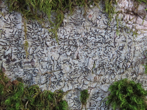
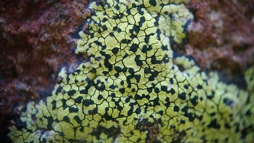
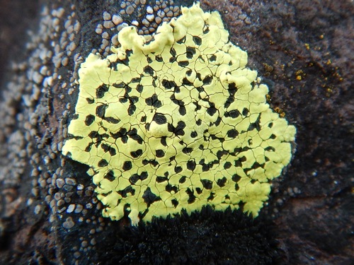 Crustose
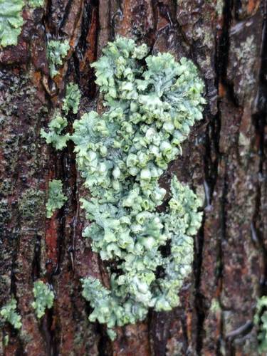
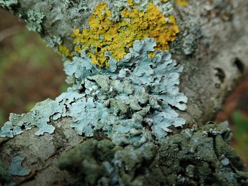
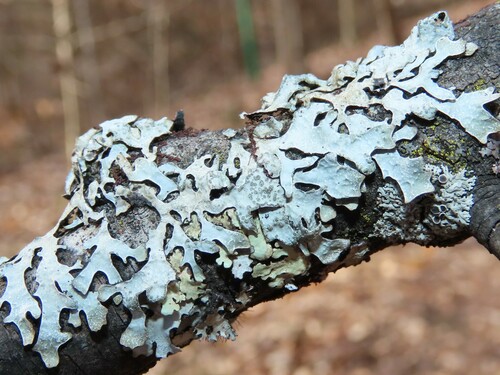
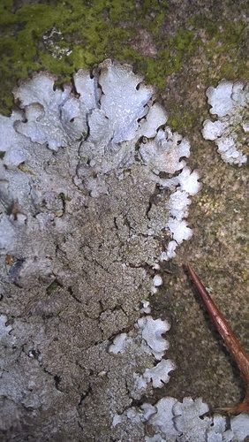
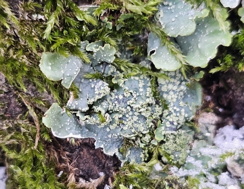
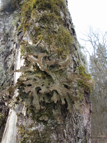 Foliose
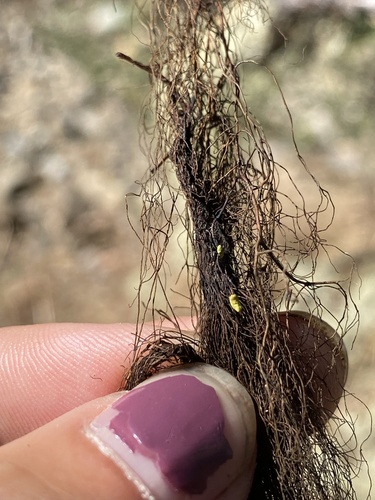
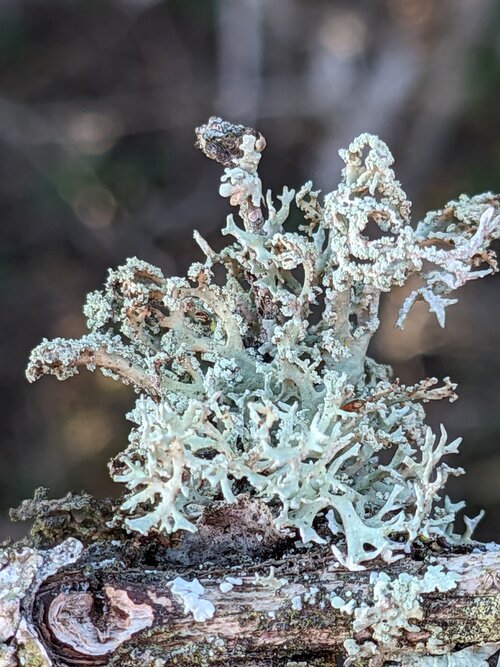
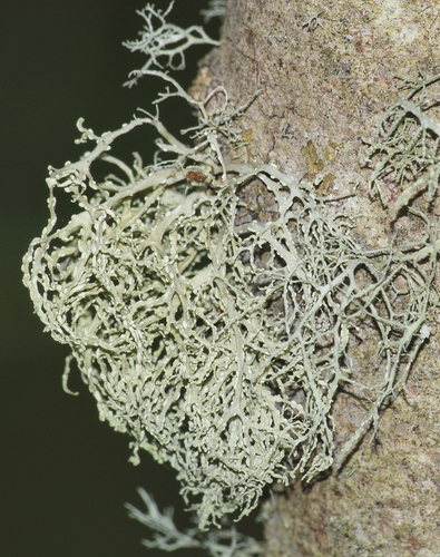
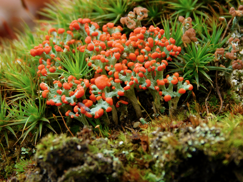
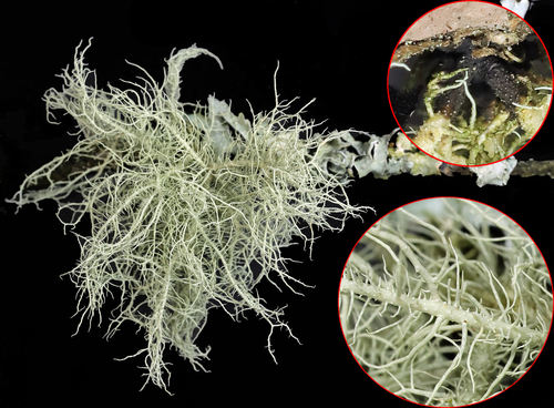 Fruticose
 Squamulose
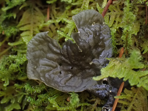
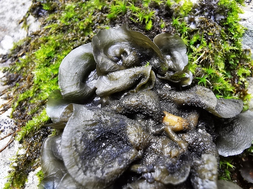
 Gelatinous
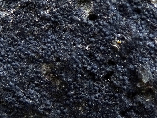
 Leprose / Crustose
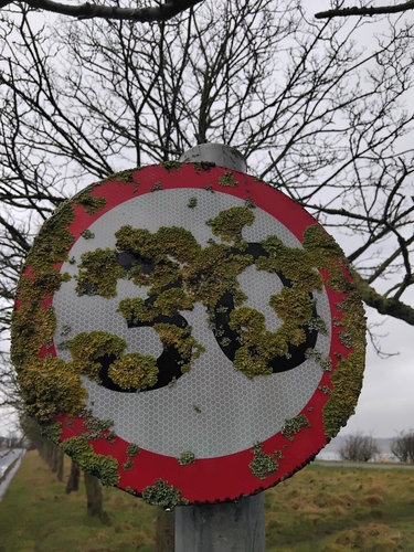
Color
Record the color carefully, and note whether the specimen is wet or dry, since many lichens change color dramatically with moisture. Dry gray-green lichens often become vivid green when wet. Bright yellows and oranges usually indicate specific chemical compounds (usnic acid, anthraquinones) that narrow identification immediately.
Substrate
What the lichen is growing on matters enormously. Many species are strict specialists: Xanthoria parietina favors nutrient-enriched bark and rock, while Rhizocarpon geographicum grows exclusively on siliceous rock. Note the specific tree species if on bark, and rock type (limestone vs. granite) if saxicolous.
 Saxicolous (rock)
 Corticolous (bark)
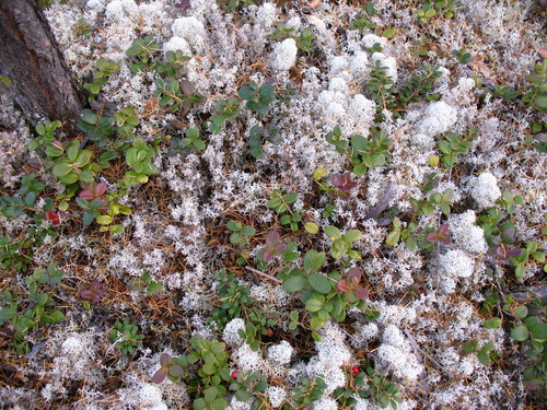 Terricolous (soil)
 Lignicolous (wood)
Lobe Shape
For foliose lichens, lobe morphology is critical. Are lobes broad (over 5 mm) or narrow? Are the tips rounded, truncate, or pointed? Do the margins curl up, lie flat, or curl under? Are the lobes tightly appressed to the substrate or loosely ascending? These details separate genera that look superficially similar.
Reproductive Structures
Look for apothecia (open disc-shaped fruiting bodies), perithecia (flask-shaped bodies embedded in the thallus), soredia (powdery granules), isidia (finger-like outgrowths), or lobules (tiny lobe-like propagules). The presence or absence of these structures is one of the most important species-level characters.
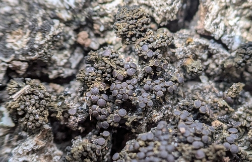
 Apothecia
 Perithecia
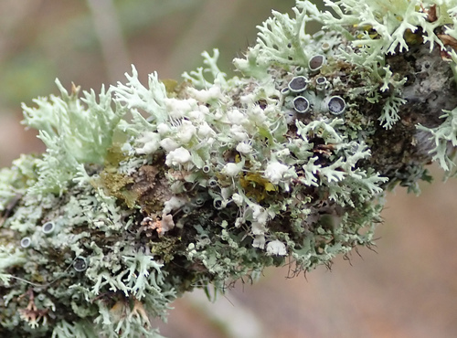
 Soredia
 Isidia
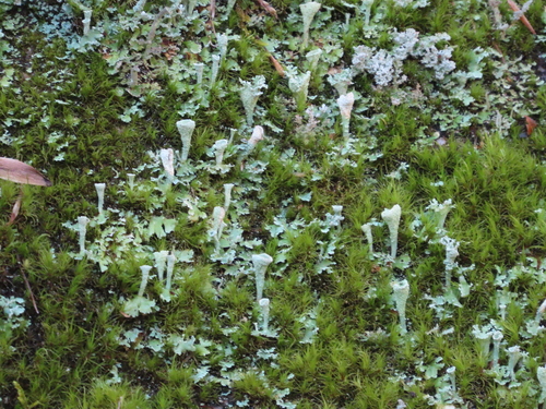
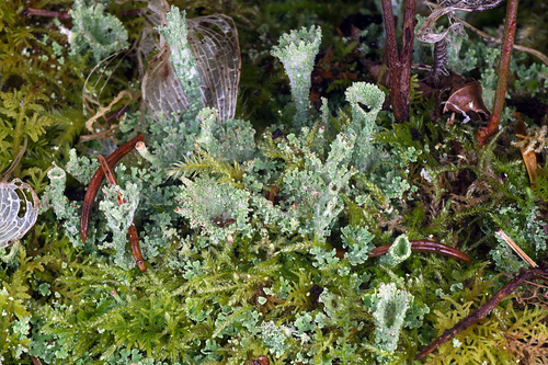
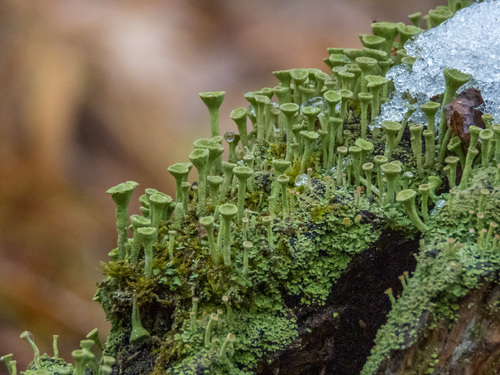
 Podetia
Underside Examination
Gently lift a lobe edge to examine the lower surface. Is it black, brown, white, or tan? Are there rhizines (root-like attachment structures), and if so, are they simple, squarrose (brush-like), or dichotomously branched? Some genera have distinctive undersurface features: Peltigera has raised veins, and Sticta has round pores called cyphellae.
 Rhizines & veins (Peltigera)
 Cyphellae (Sticta)
 Pseudocyphellae (Punctelia)
Special Features
Look for cilia (hair-like projections from lobe margins), pseudocyphellae (white dots or lines on the upper surface that are pores in the cortex), tomentum (a fuzzy felt-like covering), or pruina (a frosty white coating). These micro-characters often clinch the identification when everything else is ambiguous.
Geography & Habitat
Where you are matters. Many lichens have restricted geographic ranges, and distribution is a valid identification character. Note your region, elevation, forest type (old-growth vs. secondary), aspect (north-facing vs. south-facing), and proximity to pollution sources. A lichen that looks like Lobaria pulmonaria but grows near a highway probably is not — it requires clean air.