
Lichen Biology & Anatomy
A fungus, a photosynthetic partner, and sometimes more: how the partnership is built, how it grows and how it reproduces.
Photo: closterium_mysterium / iNaturalist, CC BYA fungus, a photosynthetic partner, and sometimes more: how the partnership is built, how it grows and how it reproduces.
Photo: closterium_mysterium / iNaturalist, CC BYPick up a lichen and you appear to be holding one thing — a crust, a leaf, a tiny shrub. But every lichen is a stable symbiotic partnership between a fungus (the mycobiont) and one or more photosynthetic organisms (the photobiont): green algae, cyanobacteria, or both. The fungus builds the body, absorbs water and minerals, and cannot photosynthesize on its own. The photobiont manufactures sugars from sunlight. Together they create an entity that neither partner could sustain alone, colonizing habitats (bare rock, desert crusts, arctic tundra) that would kill either organism in isolation.
The fungus dominates: it makes up most of the lichen’s bulk and dictates the overall form. Lichenologists name each species after its fungal partner, because the same alga can appear in dozens of visually unrelated lichens. The photobiont is the engine; the fungus is the architect.
For most of scientific history, lichens were classified as simple plants. In 1867, Swiss botanist Simon Schwendener proposed the radical idea that they were actually a symbiosis: a fungus parasitizing an alga. Leading lichenologists such as William Nylander rejected the idea, and his dual hypothesis took decades to win general acceptance. Then in 2016, everything shifted again.
The Third Partner. In 2016, Toby Spribille and colleagues published a landmark paper reporting that basidiomycete yeasts live embedded in the cortex of some macrolichens. In two Bryoria lichens that look quite different, the amount of yeast they carried correlated with one yellow toxin (vulpinic acid), but whether the yeast causes the difference is unproven. A later survey of 339 lichen species found the yeasts absent from the great majority, so their role is still open.

The mycobiont (fungus) forms the structural framework: the dense outer cortex and the airy medulla. It absorbs water and minerals from rain, fog, and dust.
The photobiont (green algae or cyanobacteria) sits in a thin layer just below the upper cortex, positioned to catch light. It manufactures all the sugars the partnership needs.
In some lichens, mainly in the family Parmeliaceae, basidiomycete yeasts (reported in 2016) also live in the cortex. Their role is unproven: they may contribute to the lichen's defensive chemistry.
The most common photobionts. Genera like Trebouxia, Trentepohlia, and Coccomyxa produce sugars efficiently through photosynthesis. A single algal species can partner with many different fungi, generating lichens that look nothing alike. The alga is a universal battery; the fungus decides the body plan.
Partners like Nostoc and Scytonema can both photosynthesize and fix atmospheric nitrogen, converting N2 gas into biologically usable ammonia. This dual capability lets cyanolichens thrive where nitrogen is scarce. They need liquid water to become active, though, so most grow in humid forests and soil crusts; the pioneers on bare rock are mainly green-algal crusts.
Tripartite lichens get the best of both worlds. Species like Lobaria pulmonaria house green algae as the primary photobiont for efficient sugar production, plus cyanobacteria sequestered in special internal structures called cephalodia. The cephalodia act as nitrogen-fixing nodules, giving the lichen access to both carbon and nitrogen from the atmosphere alone.
Slice through a foliose lichen and you find a layered architecture as deliberate as any engineered material.

Dense, tightly woven fungal tissue that acts as the lichen's skin. Transparent enough to let light reach the photobiont below, yet tough enough to shield against UV radiation, desiccation, and grazing. Often contains pigments (melanins in sun-exposed species, usnic acid creating yellow-green hues) that serve as sunscreen. In 2016, basidiomycete yeasts were found embedded here in some lichens.
The food factory. A thin band of green algal cells or cyanobacteria positioned just below the upper cortex to intercept maximum sunlight. Fungal hyphae clasp the photobiont cells through contact structures (appressoria or haustoria), through which sugars are transferred to the fungus. This is where all photosynthesis happens.
The bulkiest layer: a loose, cottony network of fungal hyphae with abundant air spaces. Its water-repellent hyphae keep these air spaces open for gas exchange (CO2 in, O2 out); water is held mainly in the cortex and in gelatinous cell walls. The medulla is usually white, which matters for identification: chemical spot tests (K, C, KC, P) are applied to medullary tissue to detect diagnostic acids. Many unique lichen compounds (about 1,000 known) are produced and deposited here.
A dense fungal layer mirroring the upper cortex, though often darker. It protects the medulla from below and anchors attachment structures. Absent in crustose lichens, which fuse directly to their substrate. In some species, the lower cortex bears cyphellae (cup-shaped pores) or pseudocyphellae (simple breaks) that facilitate gas exchange.
How the lichen holds on to its world. Rhizines are root-like bundles of hyphae in foliose lichens. An umbilicus is a single central stalk, found in umbilicate species like rock tripe (Umbilicaria). Fruticose lichens use a simple holdfast. Crustose lichens skip the question entirely: the lower medulla bonds directly to rock or bark, inseparable from its substrate.
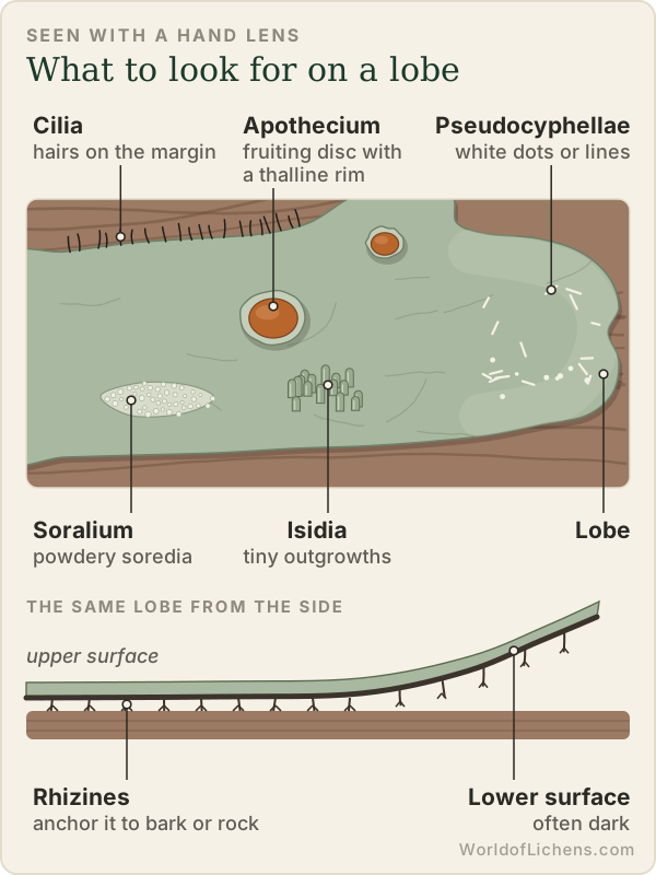
The entire lichen body. Unlike plants, it has no roots, stems, or leaves, just the layered tissue described above. The thallus can range from a thin crust a fraction of a millimeter thick to pendulous strands several meters long.
Tiny pores or breaks in the cortex that allow gas exchange. They appear as white dots, lines, or networks on the lichen surface. The distinctive white reticulate pattern on Parmelia sulcata is formed by pseudocyphellae.
Hair-like projections along lobe margins. Their color, length, density, and branching pattern are diagnostic for identification. Parmotrema species are known for conspicuous marginal cilia.
A fuzzy, felt-like covering of fine hairs, typically on the lower surface. Common in Peltigera and Lobaria. Helps trap moisture.
Updated October 2026: added how lichen structure varies between growth forms, stratified and unstratified thalli, the types of rhizine, and how to see the layers for yourself. November 2026: added diagrams of the layers in each growth form and the structures you can see with a hand lens.
The four-layer plan above describes a typical leafy (foliose) lichen. The same building blocks (a protective cortex of tightly packed fungal hyphae, a thin layer of photobiont cells, and a loose medulla) are rearranged in each growth form, and those differences are some of the most useful identification characters.
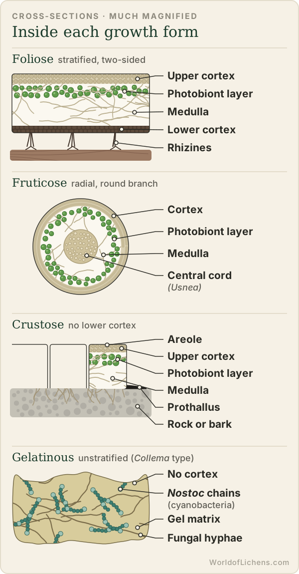
In most lichens the photobiont is confined to a distinct layer just below the upper cortex. This layered arrangement is called a stratified (heteromerous) thallus. In many gelatinous cyanobacterial lichens such as Collema, the Nostoc chains are spread evenly through the whole thallus instead, an unstratified (homoiomerous) thallus. Collema has no cortex at all, while Leptogium has a cortex only one cell thick, a difference seen clearly in a microscope section.
| Growth form | Arrangement of layers | Attachment | Example |
|---|---|---|---|
| Foliose | Upper cortex, photobiont layer, medulla, lower cortex: a flat sandwich with a distinct top and bottom (dorsiventral). | Rhizines, a felty tomentum, or a single central umbilicus | Parmelia, Umbilicaria |
| Foliose without a lower cortex | Upper cortex, photobiont layer, medulla; the underside is exposed medulla, often with raised veins. | Rhizines growing from the veins | Peltigera |
| Fruticose (round branches) | Radial: cortex on the outside, a ring of photobiont cells, then medulla. Usnea adds a dense, elastic central cord. | A basal holdfast | Usnea, Alectoria |
| Fruticose (hollow stalks) | Podetia are tubes: an outer layer (corticate, sorediate or granular) around a hollow centre. | Rise from basal squamules on soil or wood | Cladonia |
| Crustose | Upper cortex, photobiont layer and medulla, with no lower cortex; the medulla grows into the bark or rock. Many crusts are cracked into small islands (areoles) and edged by a fungal margin, the prothallus, which is black in Rhizocarpon. | The medulla itself | Rhizocarpon, Graphis |
| Leprose | No cortex and no layers: a mass of loose granules, each a few photobiont cells wrapped in hyphae. | Loosely on the surface | Lepraria |
| Gelatinous | Usually unstratified: cyanobacteria spread through a gel-rich fungal matrix that swells when wet. | Broad contact with the substrate | Collema, Leptogium |
Rhizines are bundles of fungal hyphae that grow from the lower surface and glue the lichen to its substrate. They anchor the lichen but are not roots: they do not take up water or nutrients in the way plant roots do. Their form is a standard identification character:
Other attachment types are a felty tomentum (common on Peltigera and Lobaria), the single central umbilicus of rock tripes, and the basal holdfast of fruticose lichens.
Lichens have no roots, waxy cuticle or way to hold water against drying. Their water content simply follows their surroundings, and they switch between dormancy when dry and full activity when wet. When a lichen is soaked, the cortex becomes more translucent and the green photobiont layer shows through, which is why so many grey lichens turn green after rain. Lichens with green algal partners can reactivate from humid air alone, while those with cyanobacteria generally need liquid water.
This is also why chemical spot tests are applied to a scratched surface: many diagnostic compounds sit in the medulla, not in the cortex. See the spot-test reference on the identification page.
Eight distinct body plans for conquering every surface on Earth: from paint-thin crusts fused to granite to wispy filaments trailing from branches in cloud forests.

Paint-like coatings fused directly to rock, bark, or soil. You cannot peel a crustose lichen off its substrate without destroying both. This total integration is their survival strategy: they weather extremes that would dislodge any loosely attached organism.
Crustose lichens are the most species-rich growth form and include some of the oldest living things. The largest thalli of Rhizocarpon geographicum (map lichen) have been estimated, by extrapolation, at several thousand years old. Growth rates can be agonizingly slow: often well under 0.5 mm a year, and about 0.1 mm a year in some arctic crusts.
Graphis scripta, the script lichen, belongs here too; its lirellae (elongated fruiting bodies) look like ancient writing scratched into smooth bark.

Leaf-like, with distinct upper and lower surfaces. The key test: you can peel the edges away from the substrate. Foliose lichens attach by rhizines (root-like bundles of hyphae) or, in umbilicate species, by a single central stalk.
This is one of the most familiar growth forms, ranging from a few millimetres across to rosettes 30 cm or more in diameter. Lobe shape, width, and branching pattern are the primary identification features.
Key genera: Parmelia, Flavoparmelia, Xanthoria (the bright orange sunburst lichen), and Lobaria pulmonaria (lungwort lichen, an old-growth indicator).

Three-dimensional, shrubby, or pendulous. Fruticose lichens grow upright like tiny bushes or drape from branches in long beards. They attach at a single basal holdfast. Most are radial, with cortex all round and no distinct “upper” and “lower” side; a few strap-like genera (Evernia, Pseudevernia) do have distinct upper and lower sides, and reindeer lichens have no true cortex at all.
Because they maximize surface area exposed to the atmosphere, fruticose lichens are generally the most sensitive to air pollution. Their disappearance from a forest is an early warning of declining air quality.
The genus Usnea (old man's beard) has a diagnostic elastic central cord. Pull a strand gently and it stretches. Cladonia includes the famous “British soldiers” (red-tipped podetia) and reindeer lichen. Bryoria forms dark horsehair-like tangles.
Cladonia species start as a basal mat of squamules, then develop upright podetia, hollow vertical stalks that take wildly different forms:
Cladonia pyxidata and C. chlorophaea form tidy goblet shapes. Spore-producing structures line the cup rims. Soredia may dust the insides.
Cladonia cristatella produces bright scarlet apothecia atop simple podetia, distinctive on rotting logs across eastern North America.
Cladonia rangiferina and C. arbuscula develop highly branched, antler-like podetia that form deep carpets across boreal and arctic tundra. Caribou herds depend on these mats as winter forage.
Cladonia fimbriata produces elegant flared trumpet or horn shapes, often with delicate sorediate surfaces.

Small, scale-like lobes that overlap like shingles on a roof. Squamulose lichens occupy a middle ground between crustose and foliose; their lobes are tiny and have a rudimentary upper surface, but they peel only slightly from the substrate. The basal squamules of Cladonia species are a common example. Psora forms handsome tiled crusts on calcareous soil.

Entirely powdery and granular, with no organized internal structure at all: no cortex, no layered anatomy. A leprose lichen is essentially a continuous mat of soredia-like granules. Lepraria (dust lichens) look like a dusting of pale green or blue-green powder on shaded bark or rock overhangs. They thrive in humid, sheltered microhabitats where more structured lichens cannot establish.

Rubbery and olive-green when wet, dark and brittle when dry. Gelatinous lichens use cyanobacteria (typically Nostoc) as their photobiont. Because the cyanobacterial cells are distributed throughout the thallus rather than in a neat layer, these lichens lack the structured anatomy of other types.
The transformation with moisture is dramatic: a dried, crispy black fragment can swell into a translucent, jelly-like lobe within minutes of rainfall. This is why they are often overlooked by beginners: they are nearly invisible when dry.
Key genera: Leptogium, Collema, Enchylium. All are nitrogen-fixers, making them ecologically important despite their unassuming appearance.
Two additional growth forms are less frequently encountered but complete the full spectrum of lichen body plans.
Byssoid lichens form wispy, cotton-like tufts of loosely woven fungal hyphae. They tend to occupy sheltered, humid microhabitats: the undersides of overhanging rock faces, deep bark crevices, and the interiors of hollow trees. Their loose structure maximizes moisture absorption in the still, damp air of their niche.
Because they are rarely in plain sight, byssoid lichens are easily overlooked; examples include Dictyonema and Coenogonium. When found, they resemble tiny tufts of raw cotton or spider silk tinged with green or blue-green.
Filamentous lichens consist of very fine, thread-like strands, finer even than the hair-like fruticose forms. Typical genera such as Ephebe, Cystocoleus and Racodium form tangled mats, mostly on damp or shaded rock. The photobiont cells are often arranged in chains along the filaments rather than in a discrete layer.
Like byssoid lichens, filamentous forms are easily overlooked. They may be confused with free-living algal threads or fungal mycelia until examined under magnification, which reveals the characteristic interweaving of fungal and photobiont tissues.
Not all lichens fit neatly into one category. Growth forms exist on a continuum. A lichen may be crustose at the center but foliose at the margins. Cladonia starts squamulose and becomes fruticose. Some placodioid lichens are crustose centrally with distinct lobes at the edges. In the field, use growth form as a starting point, not a rigid box.
| Form | Attachment | Remove from substrate? | 3D Structure | Key Genera |
|---|---|---|---|---|
| Crustose | Entire lower surface fused | No — destroys both | Flat | Rhizocarpon, Graphis |
| Foliose | Rhizines or umbilicus | Yes — edges peel | Leaf-like lobes | Parmelia, Xanthoria |
| Fruticose | Basal holdfast | Yes — lifts off | Shrubby or pendant | Usnea, Cladonia |
| Squamulose | Base of scales attached | Partially | Overlapping scales | Psora, Cladonia (basal) |
| Leprose | Loosely aggregated | Crumbles — powdery | None | Lepraria |
| Gelatinous | Broad contact | Yes, when wet | Rubbery lobes | Leptogium, Collema |
| Byssoid | Loose hyphae on surface | Yes — lifts easily | Cotton-like tufts | Dictyonema, Coenogonium |
| Filamentous | Tangled on substrate | Yes — peels as mat | Thread-like strands | Ephebe, Cystocoleus, Racodium |
A lichen is a partnership, but it is classified as a fungus. Each lichen species takes the scientific name of its fungal partner; the alga or cyanobacterium inside it has a separate name of its own. So Cladonia cristatella is, formally, the name of a fungus that lives with a green alga.
The standard count is about 20,000 species. The 2016 classification of lichen-forming fungi by Lücking, Hodkinson and Leavitt listed 19,387 species in 995 genera, 115 families, 39 orders and 8 classes; a 2017 correction raised it to 19,409 species.
Almost all of them, about 98%, have a fungal partner in the Ascomycota, the sac fungi, and the largest single class is the Lecanoromycetes, with over 15,000 species. Only about 170 species (under 1%) are basidiolichens, whose fungus belongs to the Basidiomycota, the group that includes mushrooms.
Lichens are not one branch of the tree of life. Fungi took up the lichen habit independently several times: a 1995 DNA study proposed at least five separate origins, and the 2016 classification estimated 20 to 30.
DNA work keeps redrawing the genera, so names change. The Elegant Sunburst Lichen was long called Xanthoria elegans; in 2003 it was moved to a new genus as Rusavskia elegans, a split that later molecular studies supported. Older field guides often still use the former names.
Here lies the central puzzle of lichen biology. A lichen must reproduce both partners together, or the fungus must find a new photobiont after dispersal. This constraint has driven the evolution of two fundamentally different strategies: sexual reproduction (fungus only, risky) and asexual reproduction (both partners together, reliable).
In sexual reproduction, only the fungal partner produces spores. These spores are shot into the wind, and if they land in the right place, they must encounter and capture a compatible free-living alga or cyanobacterium to form a new lichen. It is a gamble, but the genetic recombination it provides can be worth the risk.

The most visible reproductive structures on a lichen. Apothecia are open, cup- or disc-shaped fruiting bodies that produce fungal spores (ascospores) via meiosis. They come in two main types:
The orange discs on Xanthoria parietina and the brown shields on Peltigera are apothecia, and so, often, are the black dots on crustose rock lichens (others are perithecia or pycnidia).
Flask-shaped fruiting bodies embedded within the thallus, invisible except for a tiny opening (ostiole) at the surface. Spores are squeezed out through this pore. Less conspicuous than apothecia, perithecia are common in crustose lichens and easily overlooked. They appear as small dark dots (like pinpricks) scattered across the thallus surface.
Elongated, slit-like fruiting bodies that look like lines of ancient script written on bark. They are the defining feature of the aptly named Graphis scripta (script lichen). Lirellae can be straight, curved, branched, or stellate, and their morphology is important for identification within the Graphidaceae family.
Asexual reproduction sidesteps the fundamental gamble of sexual reproduction. Instead of dispersing naked fungal spores that must find a new photobiont, the lichen packages both partners together in a single dispersal unit. No partner-finding required. The tradeoff: no genetic recombination.
One of the most important distinctions in lichen identification. Two mechanisms that solve the same problem (vegetative dispersal) in fundamentally different ways.

Tiny powdery granules, each a few algal cells wrapped in a wisp of fungal hyphae. No cortex. They feel granular to the touch. Produced in structures called soralia, where the lichen's cortex breaks open to release them.
Soralia placement is diagnostic: they can be laminal (on the upper surface), marginal (along lobe edges), or apical (at lobe tips). Parmelia sulcata has soredia erupting from cracks in its surface network.
Term to remember: a lichen with soredia is called “sorediate”

Small, finger-like or coral-like outgrowths from the thallus surface. Corticated: they have a cortex layer covering them, giving them a smooth, shiny surface. They snap off and grow into new lichens.
Isidia come in various shapes: cylindrical, globular, coralloid (branching like coral), or scale-like. Pseudevernia furfuracea bears abundant coralloid isidia. Species like Parmelia saxatilis are richly isidiate.
Term to remember: a lichen with isidia is called “isidiate”
Why does this matter so much? Many lichen species pairs are distinguished only by whether they produce soredia or isidia. Parmelia sulcata (sorediate) vs. Parmelia saxatilis (isidiate). Same genus, similar habitat, nearly identical appearance, until you reach for a hand lens and check the surface texture. Powdery granules = soredia. Shiny fingers = isidia. That distinction can determine the species.
Miniature lobes that develop along the margins or on the upper surface of foliose lichens. Larger and more structured than isidia, lobules are essentially small copies of the parent thallus. They break off and establish independently.
The simplest method of all. A piece of the thallus breaks off (snapped by wind, carried by a bird, washed by rain) and grows into a new individual wherever it lands. This is especially common in fruticose lichens. Storm-blown fragments of Usnea on the forest floor can successfully re-establish on new branches.
Sexual reproduction is a lottery. Asexual reproduction is a sure thing, but at the cost of genetic diversity. Many lichens hedge their bets and do both.
How lichens build soil from bare rock, monitor air pollution, and survive conditions that would kill nearly any other organism on Earth.